Craniopharyngiomas

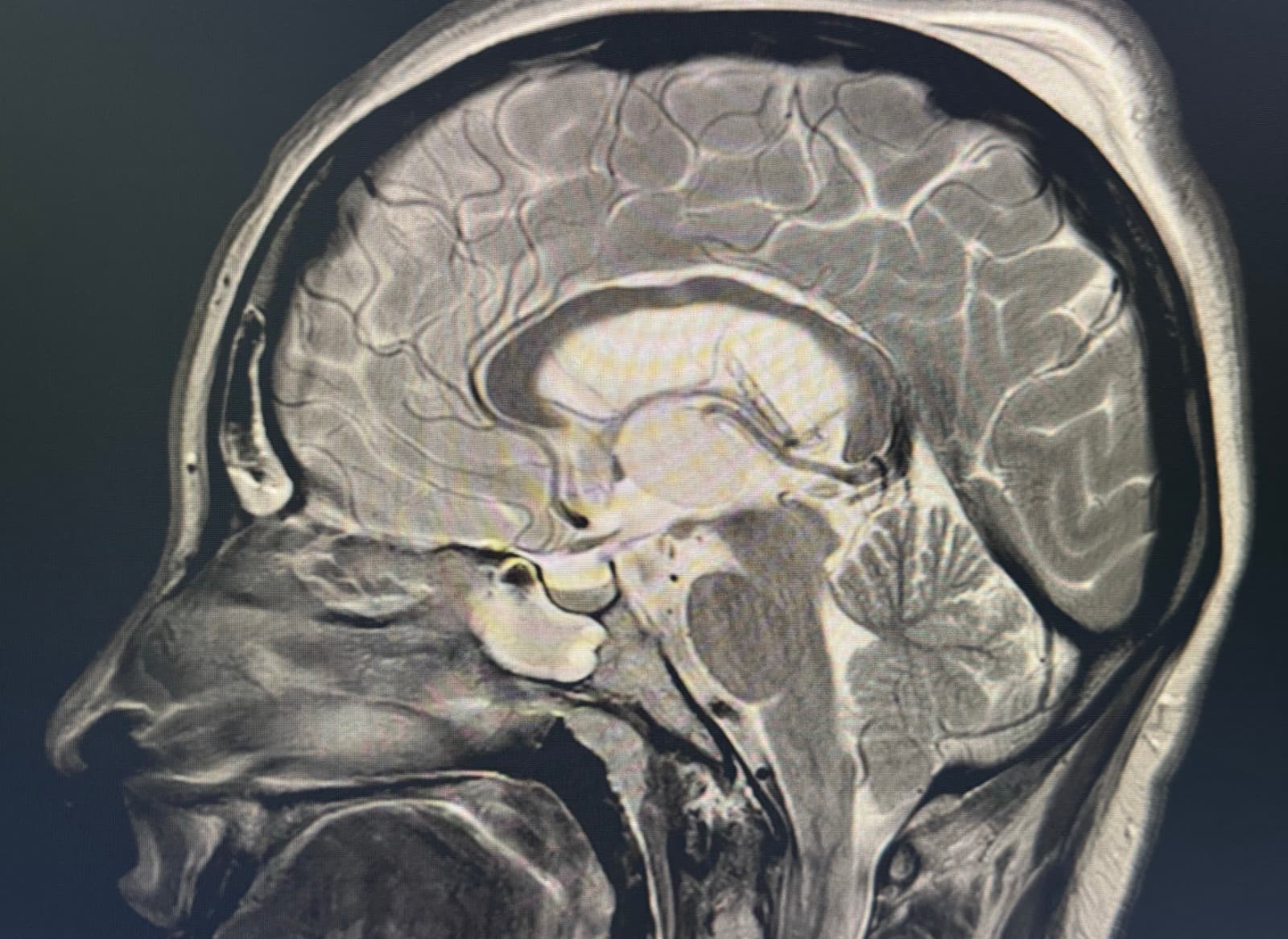
Craniopharyngiomas are benign tumors of the sellar and suprasellar region derived from epithelial cells of Rathke's pouch. They have a higher incidence in children and older adults. The most common symptoms are visual field alterations, hormonal deficits, and short stature in children. When these tumors cause hydrocephalus, they may present with headache, vomiting and altered consciousness.

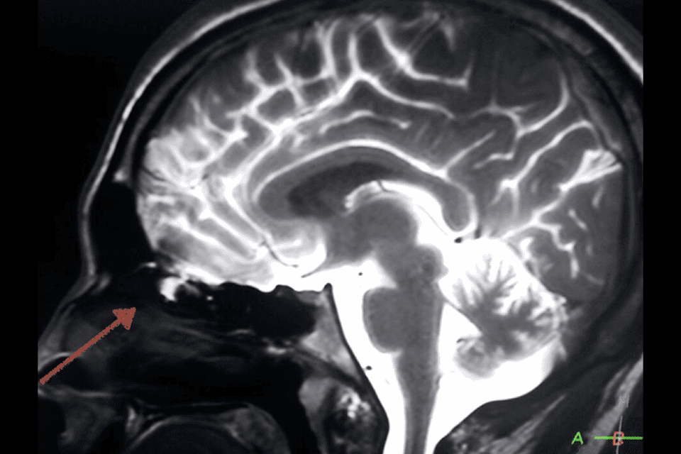
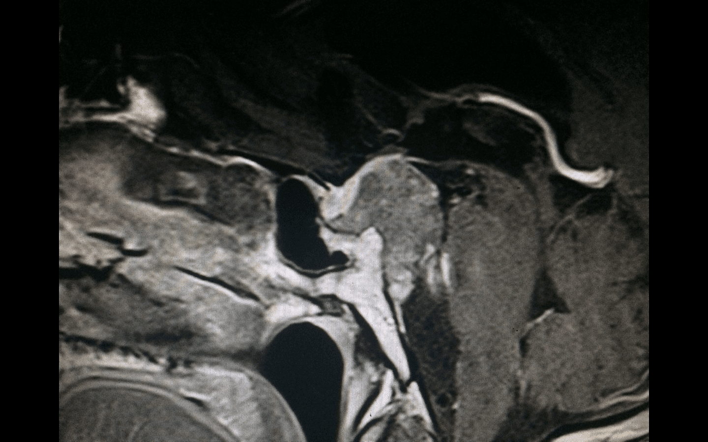
We present the case of a 20-year-old patient who consulted for progressive visual deficit accompanied by drowsiness due to cortisol deficiency. In this case we were able to perform a complete resection of the craniopharyngioma; the postoperative course was uneventful, complete hormonal replacement was carried out, and we did not need to use radiotherapy.

Note the high definition of the intraoperative images provided by the endoscopic endonasal approach. This visualization allows us to make the most appropriate decisions during surgery to deliver a good outcome.
Symptoms

  • Visual field alterations
  • Hormonal deficits
  • Short stature in children
  • Headache, vomiting and altered consciousness when the tumor causes hydrocephalus

Diagnosis

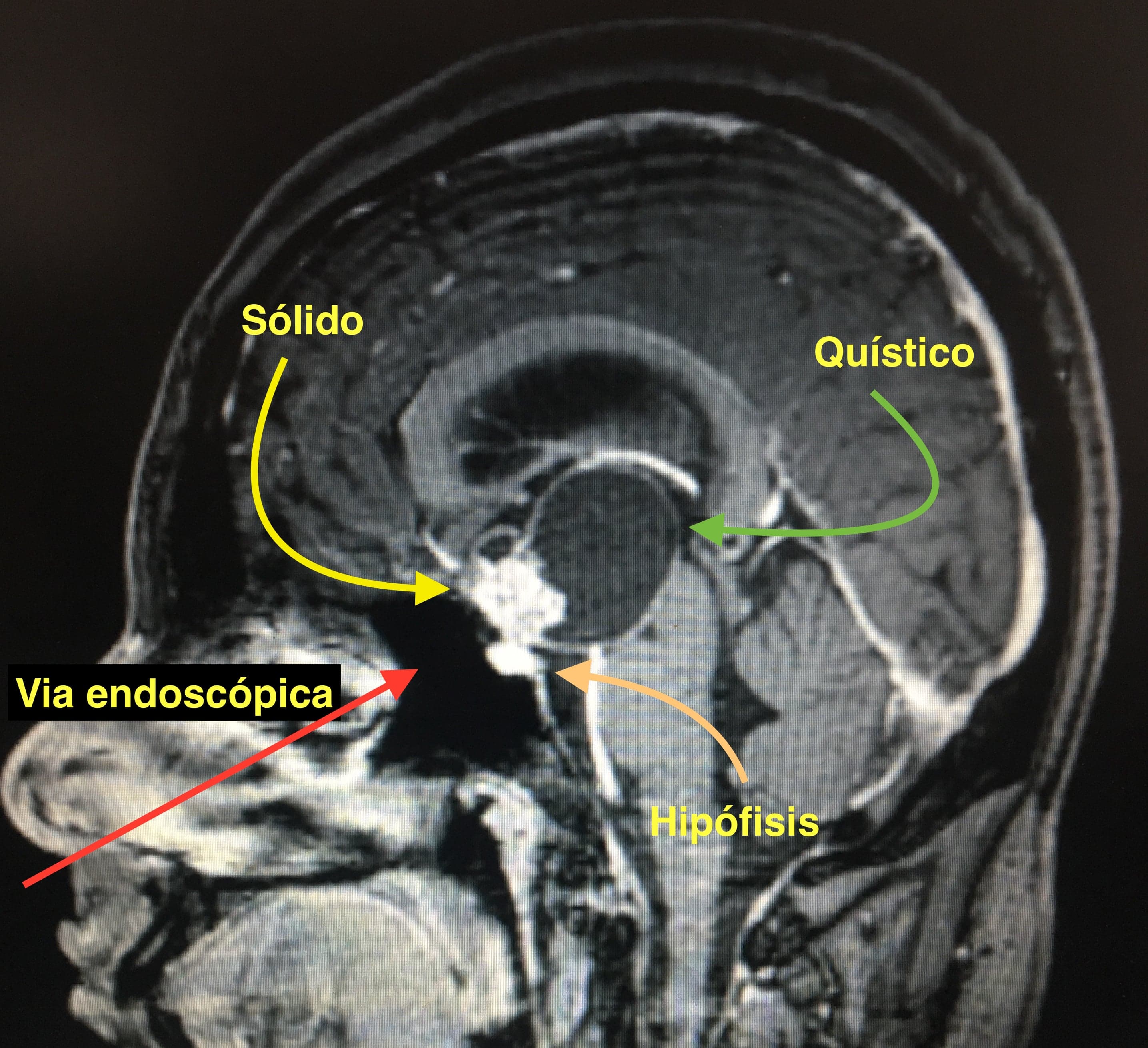
  • Magnetic resonance imaging (MRI) — shows the typical solid-cystic suprasellar tumor with calcifications
  • Brain computed tomography (CT) — useful to assess the calcifications and ventricular size

Treatment

The best treatment for these tumors is complete resection when possible. We have radiotherapy as adjuvant therapy for active tumor remnants or for recurrent tumors.

Because craniopharyngiomas originate in the hypothalamic-pituitary axis, are primarily midline and are in intimate relationship with the skull base, the endoscopic endonasal approach is the most commonly chosen pathway, as it is the most direct to the region.

Our approach

  • Prioritization of the endoscopic endonasal route, the most direct to the region
  • Better visualization of the tumor and its relationship with the hypothalamic-pituitary axis
  • Goal of complete resection while preserving endocrine and visual function
  • Personalized long-term follow-up with imaging controls
Clinical case

Frequently asked questions

Do you need a consultation?

If you'd like to discuss your case or get a second opinion, you can schedule an in-person or virtual consultation.

Related pathologies

Cerebrospinal Fluid Leaks

Cerebrospinal Fluid Leaks

Skull base defects that allow cerebrospinal fluid to escape through the nasal cavities, treatable with endoscopic endonasal surgery.

View pathology
Chordomas

Chordomas

Rare, locally aggressive tumors derived from embryonic remnants of the notochord, typically located at the skull base and sacrum.

View pathology
Colloid Cyst of the Third Ventricle

Colloid Cyst of the Third Ventricle

The colloid cyst is a benign lesion typically located in the third ventricle, near the foramina of Monro — a key region for cerebrospinal fluid circulation.

View pathology