Colloid Cyst of the Third Ventricle

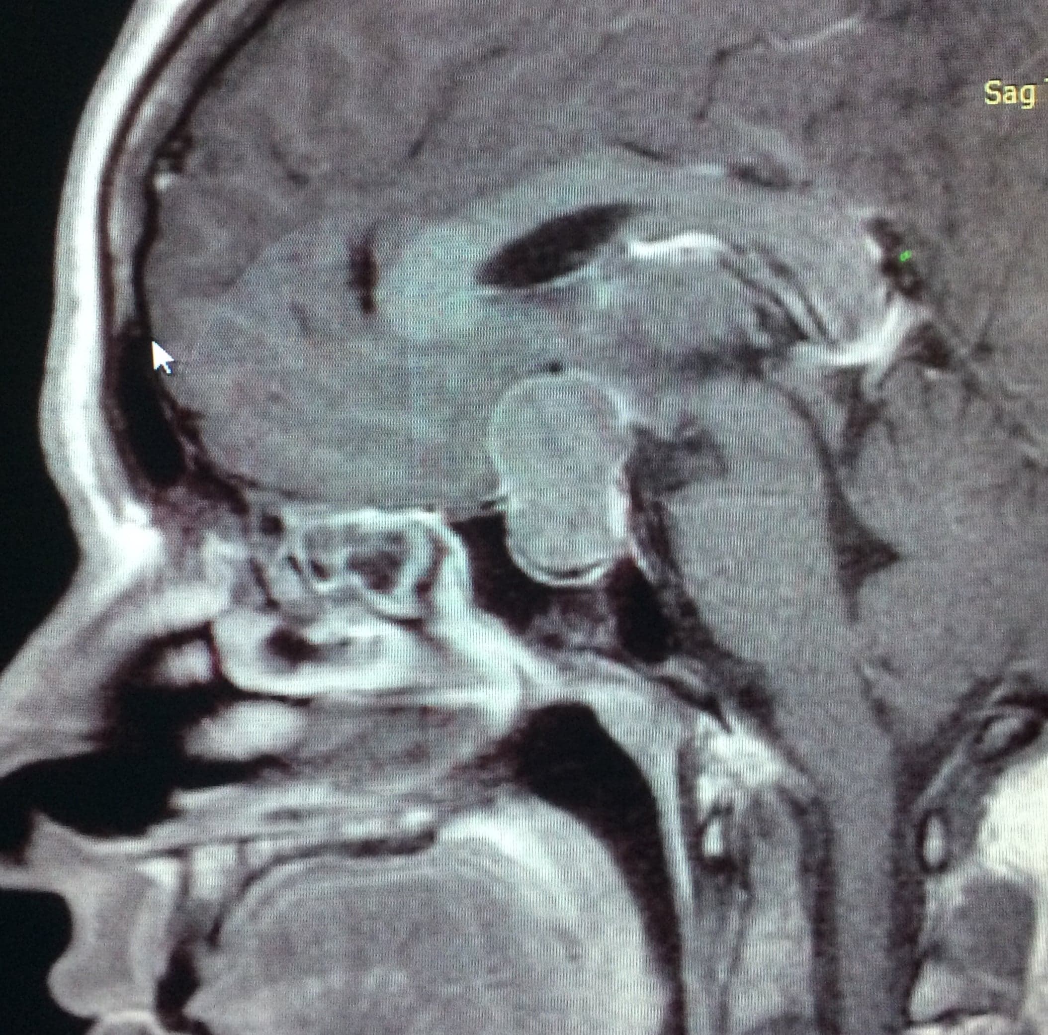
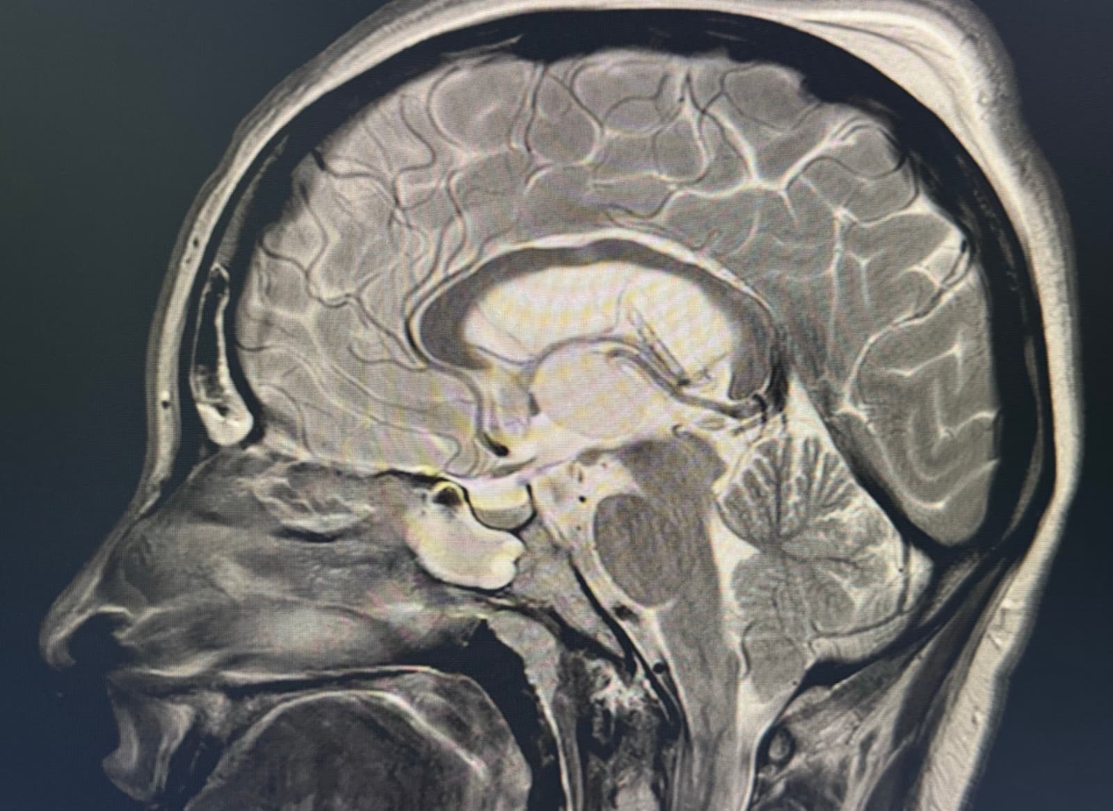
The colloid cyst is a benign lesion typically located in the third ventricle, near the foramina of Monro — a key region for cerebrospinal fluid circulation. Although it is a benign tumor, it can obstruct fluid flow and produce hydrocephalus, severe headaches, memory disturbances, visual alterations, and even serious neurological symptoms.

Our team specializes in ventricular endoscopic surgery, a minimally invasive technique that allows us to resect this type of lesion while avoiding extensive craniotomies and minimizing surgical trauma.

The clinical case we present is that of a patient with progressive headaches and episodes of intracranial hypertension. Studies showed a colloid cyst of the third ventricle associated with ventricular dilation. A complete endoscopic resection was performed through ventricular neuroendoscopy, and the patient progressed favorably, with resolution of symptoms and adequate ventricular circulation in subsequent follow-ups.

The surgery was carried out through a neuroendoscopic ventricular approach via a small incision and a millimeter-sized burr hole. Using high-definition endoscopic vision, the colloid cyst was identified, its capsule was opened, its content evacuated, and the cyst wall subsequently resected microsurgically.

This type of approach allows us to avoid extensive craniotomies, reduce brain tissue injury, achieve a faster postoperative recovery with less pain, and shorten hospitalization time.
Symptoms

  • Intense headaches, often positional or sudden in onset
  • Nausea and vomiting
  • Recent-memory disturbances and difficulty concentrating
  • Episodes of sudden loss of strength in the legs ("drop attacks")
  • Blurred or double vision
  • Dizziness and balance disorders

Diagnosis

  • Brain magnetic resonance imaging (MRI) — the study of choice, confirms the diagnosis
  • Brain computed tomography (CT) — useful in acute symptoms and to assess ventricular size
  • Complete neurological evaluation
  • Ophthalmologic evaluation with visual field testing if visual symptoms are present

Treatment

Treatment of a colloid cyst depends on the size of the lesion, the presence of hydrocephalus, and the patient's symptoms. For small, asymptomatic cysts found incidentally, periodic MRI follow-up may be indicated.

When the cyst produces symptoms, grows progressively, or causes obstruction of cerebrospinal fluid flow, surgery is the treatment of choice. The preferred technique today is ventricular neuroendoscopy, which allows the cyst to be resected through a small opening in the skull, avoiding extensive craniotomies.

In specific cases where hydrocephalus is already decompensated or endoscopic access is limited, placement of a ventriculoperitoneal shunt or a complementary microsurgical approach may be required.

Our approach

  • Team specialized in high-complexity ventricular endoscopic surgery
  • Minimally invasive approach that reduces trauma to brain tissue
  • High-definition endoscopic visualization throughout the procedure
  • Personalized long-term follow-up with imaging controls
Clinical case

Frequently asked questions

Do you need a consultation?

If you'd like to discuss your case or get a second opinion, you can schedule an in-person or virtual consultation.

Related pathologies

Esthesioneuroblastoma or Olfactory Neuroblastoma

Esthesioneuroblastoma or Olfactory Neuroblastoma

Esthesioneuroblastoma is a rare malignant tumor of the anterior skull base originating from olfactory cells.

View pathology
Pituitary Adenomas

Pituitary Adenomas

Benign, slow-growing tumors originating from cells of the adenohypophysis, classified as functioning or non-functioning.

View pathology
Craniopharyngiomas

Craniopharyngiomas

Benign tumors of the sellar and suprasellar region derived from epithelial cells of Rathke's pouch.

View pathology