Cerebrospinal Fluid Leaks

CSF leaks through the nasal cavities can be post-traumatic, following skull base surgery, or simply spontaneous — the latter being the most common cause. They result from a defect in the skull base, both in the bony and dural planes, that allows CSF to escape outward. With the brain having a communication to the exterior, there is a risk of ascending bacterial meningitis from common nasal cavity organisms.

Clinically they present as loss of a transparent water-like fluid — non-viscous, unilateral, odorless, with a salty taste — and the flow increases when the patient sits up and leans the head forward. They can be continuous or intermittent, present with recurrent meningitis, or with seizures when associated with brain tissue herniated through the defect, forming a meningoencephalocele.

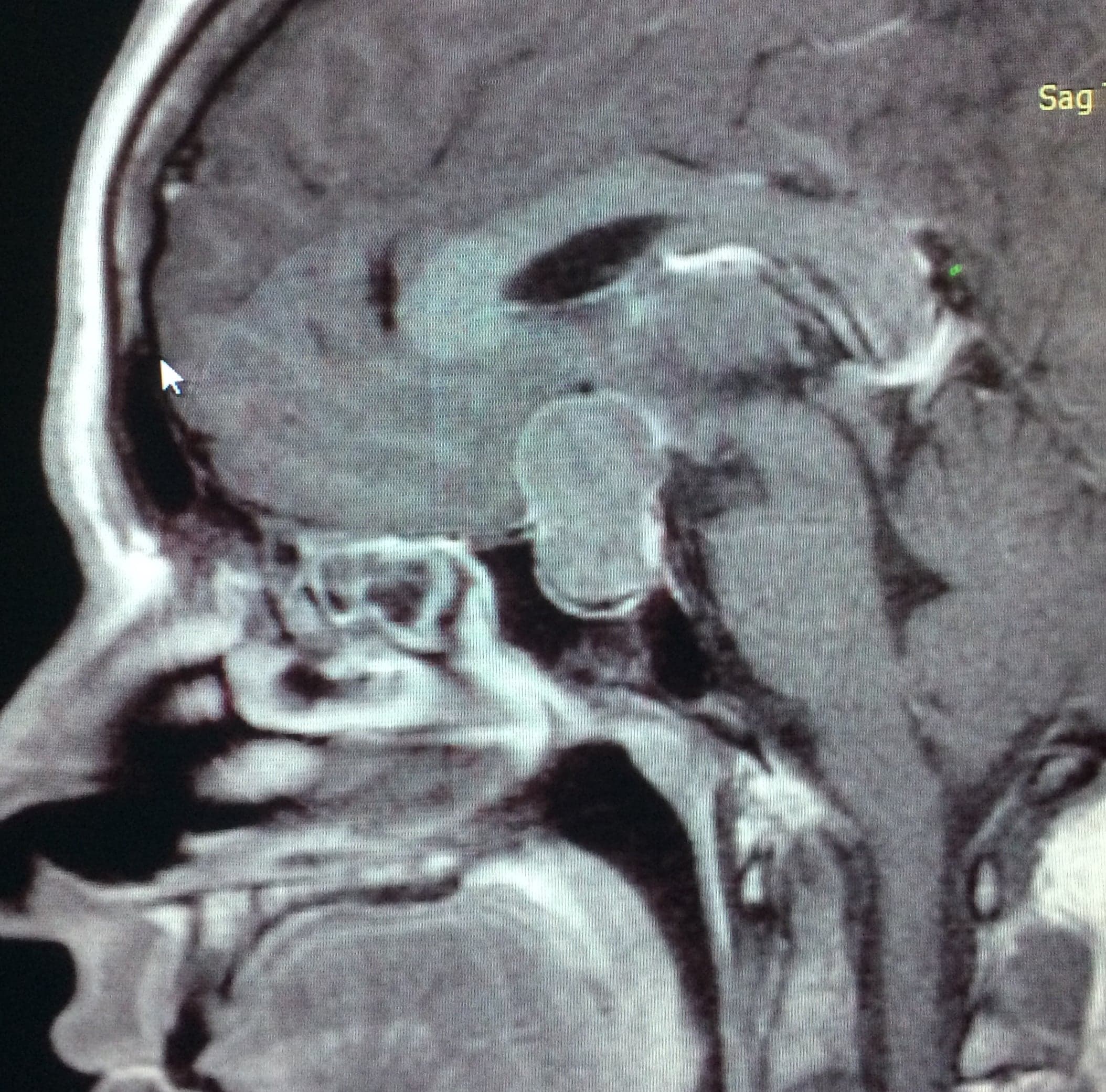
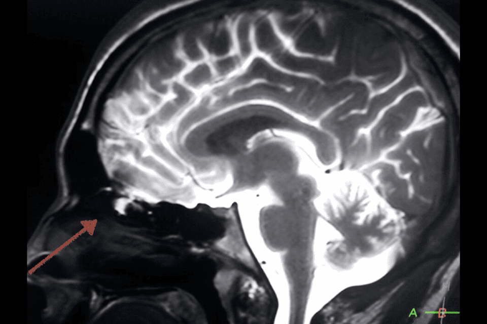
Imaging studies to localize the defect are multiaxial CT and MRI. The most frequently affected sites, in decreasing order, are the ethmoid, sphenoid sinus, frontal sinus, and lastly the clivus.

With the development of endoscopic endonasal surgery, this is currently the access route of choice for the treatment of this pathology, without the need for craniotomy. The surgery involves localizing the dural defect and interposing multiple layers of tissue to seal the fistulous tract, providing vascularization with a pediculated nasoseptal flap.
First Case

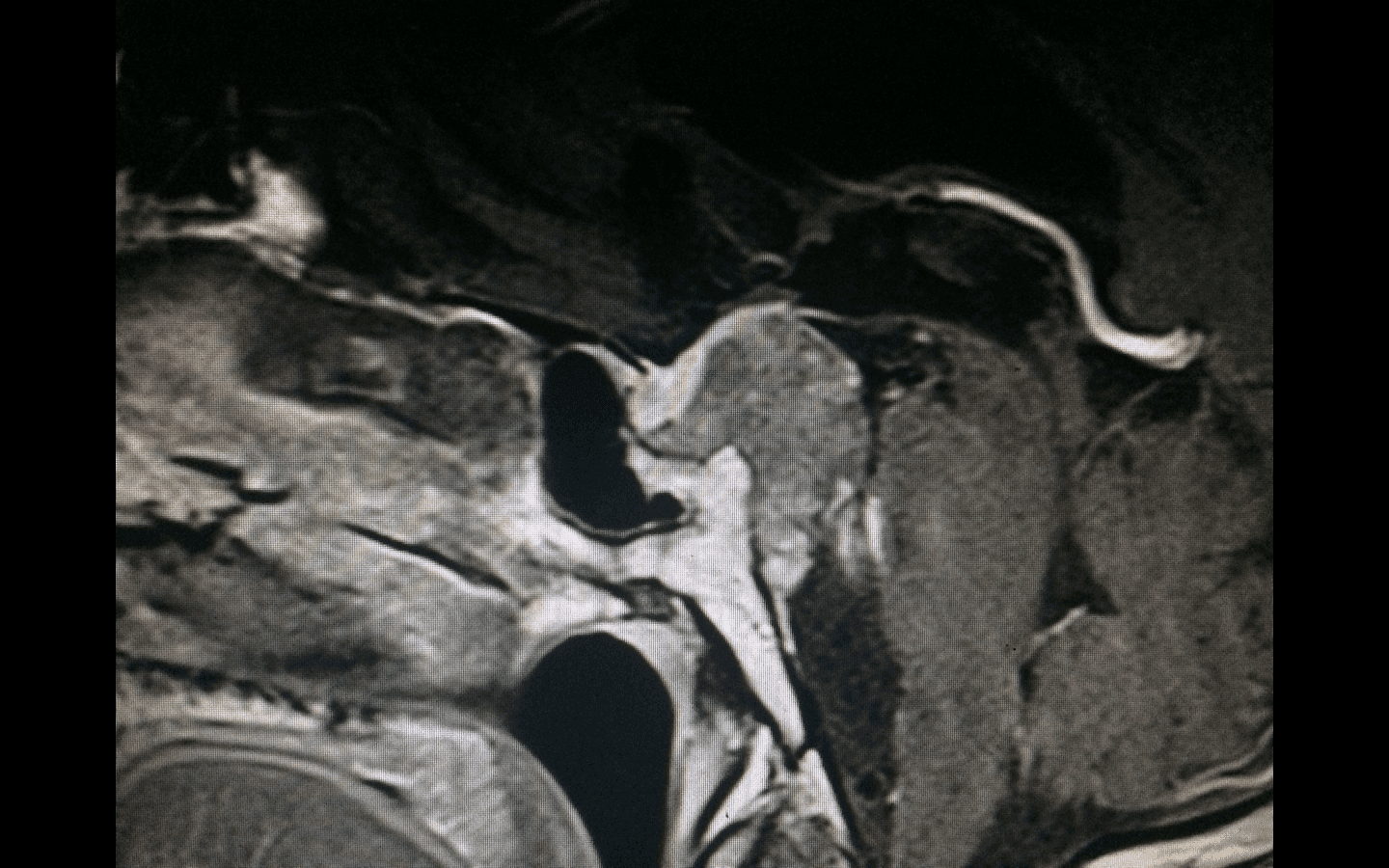
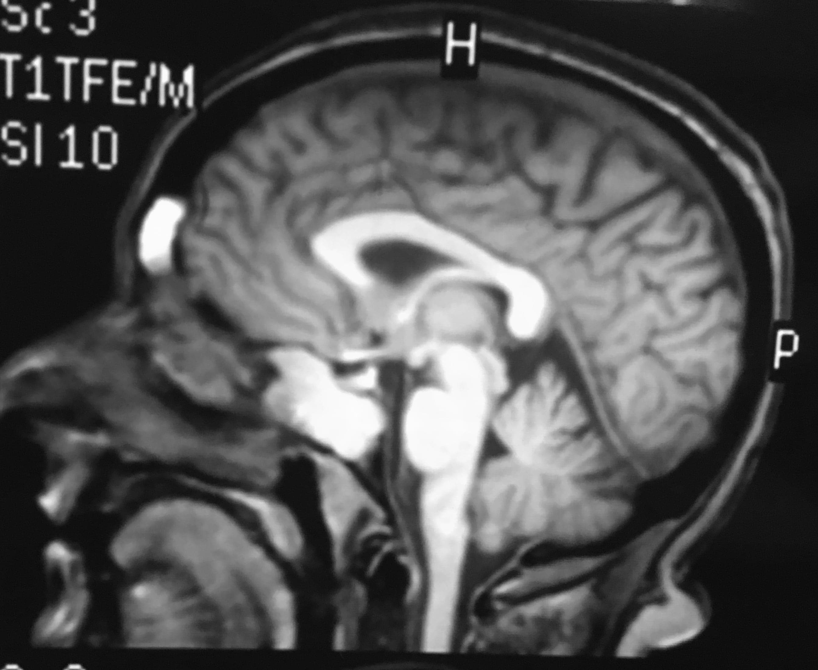
A 45-year-old patient on chronic treatment for rhinorrhea is diagnosed with a CSF leak through the nasal cavities. Imaging studies do not show the defect in the skull base, so endoscopic endonasal surgery is performed directly to localize the fistulous site and seal it. A small bony and dural defect with spontaneous CSF leakage is located in the roof of the right ethmoid — the most frequently observed site due to the natural weakness of the region. An intracranial dural substitute is interposed, and the defect is then covered with a nasoseptal flap providing vascularization to the skull base reconstruction.

Second Case

A 57-year-old patient with intermittent right-sided CSF loss associated with headache. MRI shows herniation of part of the right temporal lobe toward the sphenoid sinus. Axial CT of the skull base shows a bony defect in the right lateral aspect of the sphenoid, through which the meningoencephalocele responsible for the CSF leak herniates.

Third Case

A 22-year-old patient consults for generalized seizures with no history of fluid loss through the nasal cavities. Imaging studies show a voluminous defect in the left middle cranial fossa with an encephalocele responsible for the seizures. Endoscopic endonasal surgery is performed to resect the meningoencephalocele and reconstruct the skull base. Herniated brain tissue is visualized through a congenital defect in the skull base lateral to the maxillary nerve — known as Sternberg's Canal. Reconstruction is done with a dural substitute and a contralateral nasoseptal flap.

Related cases

Chordomas

Chordomas

Rare, locally aggressive tumors derived from embryonic remnants of the notochord, typically located at the skull base and sacrum.

View clinical case
Esthesioneuroblastoma or Olfactory Neuroblastoma

Esthesioneuroblastoma or Olfactory Neuroblastoma

Esthesioneuroblastoma is a rare malignant tumor of the anterior skull base originating from olfactory cells.

View clinical case
Pituitary Adenomas

Pituitary Adenomas

Benign, slow-growing tumors originating from cells of the adenohypophysis, classified as functioning or non-functioning.

View clinical case