
Pituitary adenomas are benign, slow-growing tumors originating from cells of the adenohypophysis. We classify them as «Non-Functioning» when they don't secrete any hormone, and as «Functioning» when they secrete biologically active hormones.
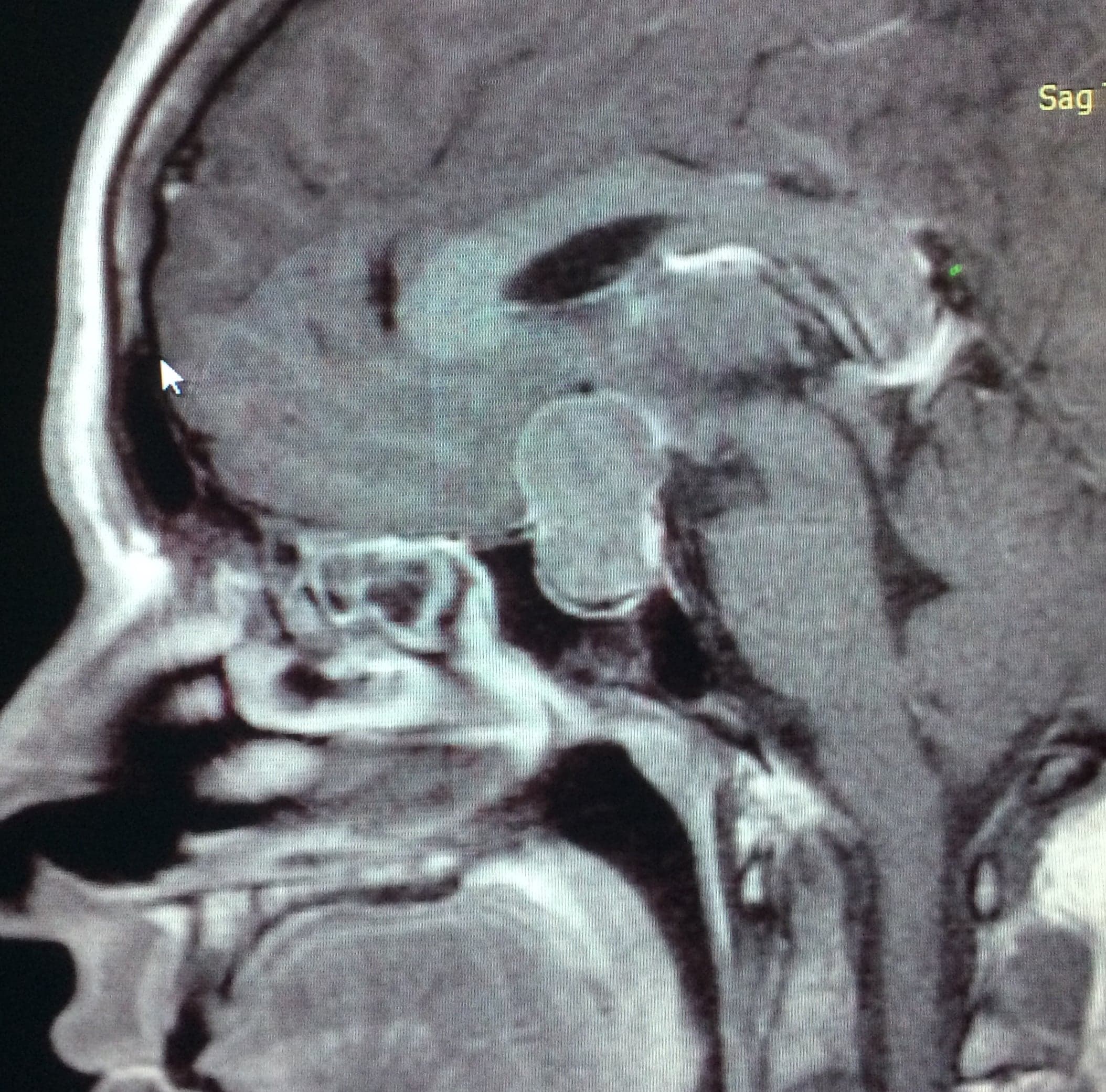
Pituitary Adenomas
Non-functioning adenoma
«Non-Functioning» tumors cause symptoms due to mass effect on adjacent structures, such as visual deficits and double vision. «Functioning» tumors are responsible for Acromegaly (from increased growth hormone) and Cushing's disease (from excess cortisol).
The first-line treatment for both functioning and non-functioning tumors is endoscopic endonasal surgery, except for Prolactinomas, where medical therapy is the first option.
The development of the endoscopic endonasal technique allows the surgical treatment to be performed with excellent visualization of the skull base, improving the limits of resection.
Cushing's Disease
Cushing's disease is caused by a pituitary adenoma that stimulates the hypersecretion of endogenous cortisol. The clinical syndrome is typically characterized by central obesity, a rounded face, muscle weakness, arterial hypertension, diabetes, and reddish-purple stretch marks. The endocrinological diagnosis is based on specific laboratory tests, and the pituitary microadenoma is usually detected on MRI. The first-line treatment is surgical, with hormonal therapy and radiosurgery as second-line options.
High-quality MRI imaging allows us to identify small microadenomas, and thanks to the excellent visualization provided by the endoscopic surgical technique, we can perform effective and precise resections.
Acromegaly
Acromegaly is a disease caused by a pituitary adenoma that excessively secretes Growth Hormone and stimulates the increase of peripheral growth factors such as IGF-1.
Clinically, excessive tissue growth is observed, along with enlarged hands and feet, increased tongue volume and characteristic facial features. Patients complain of joint pain, bilateral carpal tunnel syndrome, sweating, headache and visual disturbances. Significant metabolic changes are also present, such as type 2 diabetes and arterial hypertension.
The endocrinological diagnosis is performed through measurement of Growth Hormone, IGF-1, and the Oral Glucose Tolerance Test. We then perform MRI to identify the pituitary adenoma responsible for the disease.
The best treatment we can offer is surgical resection — depending on the location of the adenoma, we evaluate the chance of biochemical cure. We also have hormonal therapies and radiosurgery for residual, recurrent or aggressive tumors.
Pituitary Apoplexy
This term refers to ischemic or hemorrhagic infarction of the pituitary gland, presenting with acute endocrinological and neurological symptoms. Destruction of the gland causes hormonal alterations — cortisol deficiency being the most urgent to replace. The rapid increase in sellar content compresses the optic pathway and cavernous sinus, leading to decreased visual acuity and double vision. When the neurological deficit develops suddenly and progressively, surgical decompression can rapidly improve the compressive symptoms.
We can illustrate with the case of a patient on chronic anticoagulant therapy for a cardiac condition, who suffered a sudden headache accompanied by decreased visual acuity and double vision.
Invasive Adenomas
Pituitary adenomas are, for the most part, tumors that develop in the sella turcica and extend to the suprasellar cistern while respecting normal anatomical barriers. In a smaller percentage, we find adenomas that invade neighboring structures without respecting regional limits. These tumors begin by destroying the dural and bony planes of the skull base, becoming locally aggressive tumors. The most frequently affected sites are: cavernous sinus, sphenoid sinus, clivus, anterior skull base, orbital cone, nasal cavities, ethmoid sinuses, middle cranial fossa, infratemporal fossa, etc.
Clinically, they present with hormonal deficits, symptoms from neural compression such as visual deficit, double vision, and eyelid ptosis; symptoms from mass effect such as nasal obstruction, epistaxis, hyposmia; or invasive symptoms such as cerebrospinal fluid leak.
Imaging studies show skull base tumors invading the aforementioned regions, requiring differential diagnosis with aggressive skull base tumors such as undifferentiated carcinomas, esthesioneuroblastomas, rhabdomyosarcomas, lymphomas, neuroendocrine carcinoma, giant cell bone tumors, etc.
The treatment of choice for these tumors, despite their large volume, is endoscopic endonasal resection, as it allows us to reach all of the mentioned regions through a single access, without the need for additional open surgery.
Giant Adenomas
Pituitary adenomas are, for the most part, benign slow-growing tumors. The gradual increase in tumor volume causes neural structures to slowly lateralize, potentially reaching diameters greater than 4 cm — at which point they are called Giant Adenomas.
Clinically, they present with hormonal axis alterations and mass-effect symptoms such as decreased visual acuity, hemianopsia, amaurosis, double vision, hydrocephalus, etc.
Today, even when adenomas are giant, the treatment of choice in over 98% of cases is the extended endoscopic endonasal approach. These naturally require a wider approach to the skull base, extensive resection of the voluminous lesion, and reconstruction of the base due to the space left after resection. The endoscopic endonasal approach remains the safest when compared to open craniotomies, since the brain is not directly exposed and the resection of the voluminous tumor is performed without mobilization of brain tissue.
We can illustrate with the case of a 56-year-old patient with panhypopituitarism and a marked decrease in visual acuity, diagnosed with a giant pituitary tumor.
Related cases
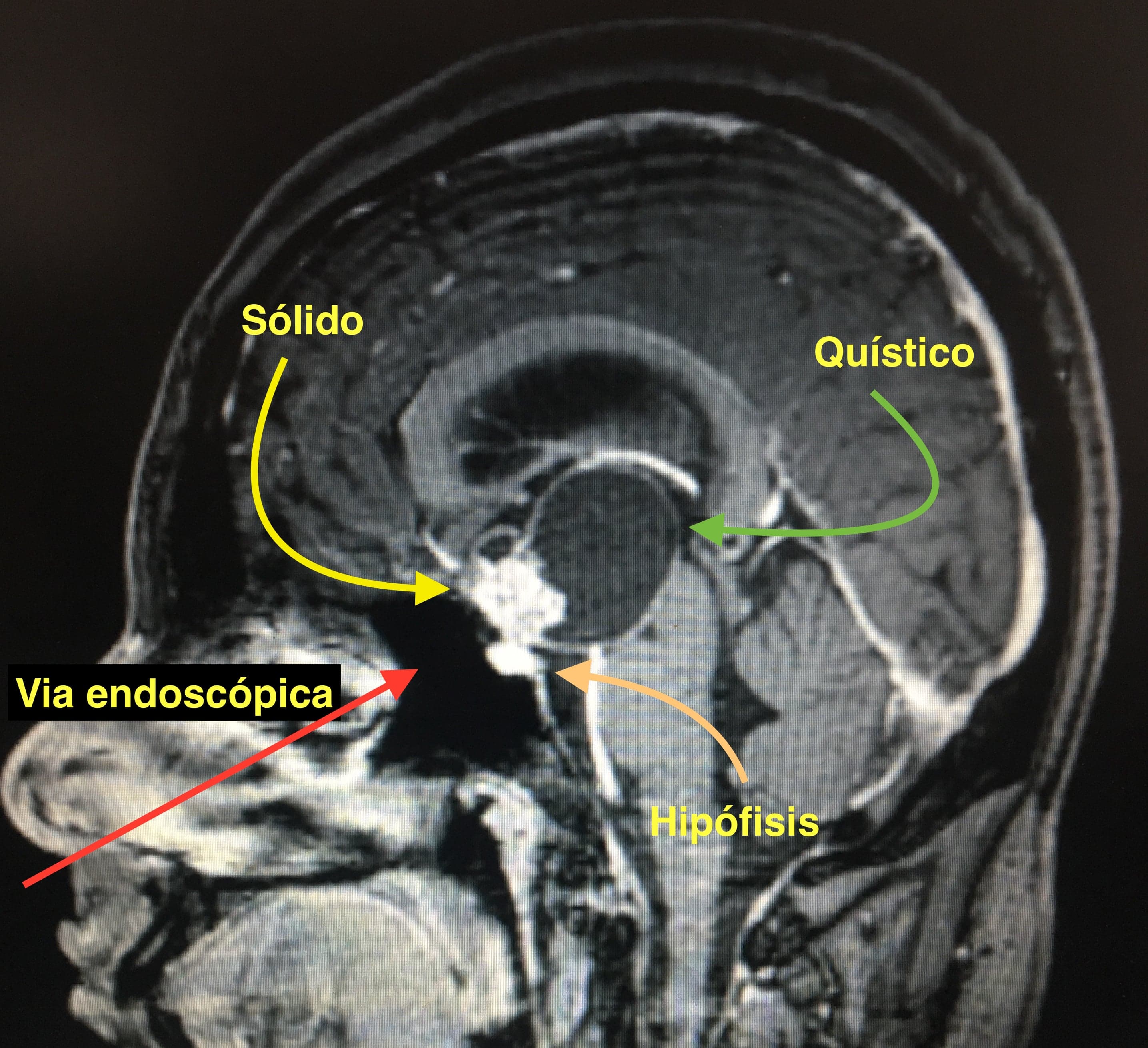
Craniopharyngiomas
Benign tumors of the sellar and suprasellar region derived from epithelial cells of Rathke's pouch.
View clinical case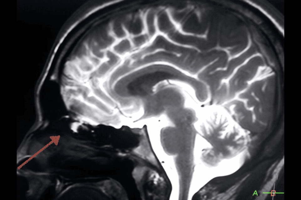
Cerebrospinal Fluid Leaks
Skull base defects that allow cerebrospinal fluid to escape through the nasal cavities, treatable with endoscopic endonasal surgery.
View clinical case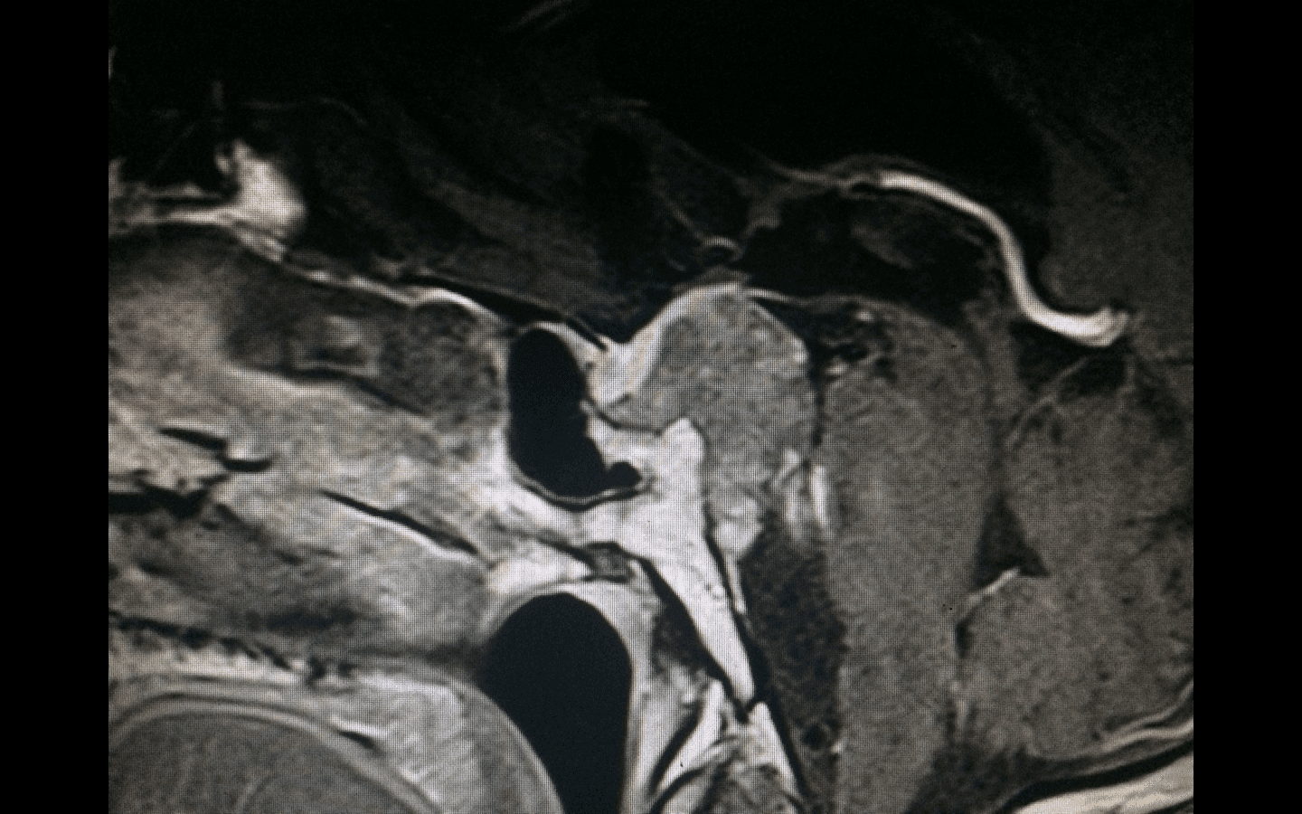
Chordomas
Rare, locally aggressive tumors derived from embryonic remnants of the notochord, typically located at the skull base and sacrum.
View clinical case