Chordomas

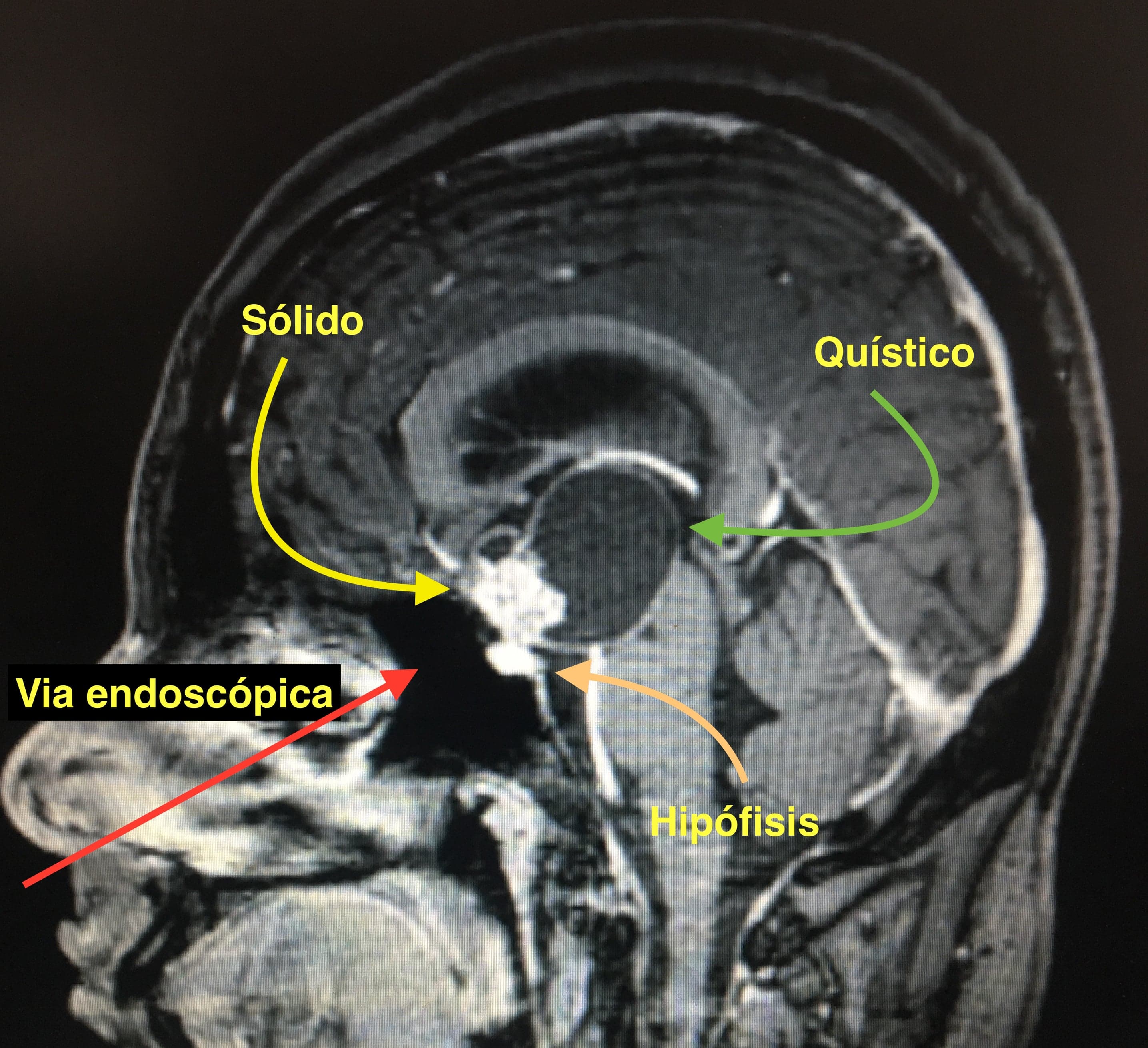
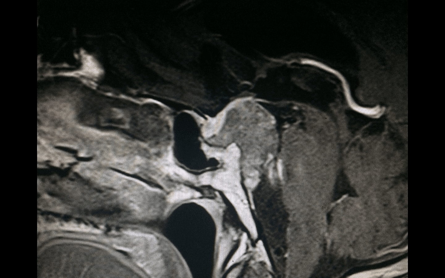
Chordomas are rare tumors arising from embryonic remnants of the notochord. They are locally aggressive, recurrent tumors that invade both bony and soft tissue. The biological nature of chordomas makes them midline central tumors, most commonly located at the skull base and the sacrum. Symptoms depend on location — in this description we will focus on those at the skull base.

They originate in the bony tissue of the clivus, and their growth can extend either outward — toward the paranasal sinuses, pharynx, paravertebral muscles or the first cervical vertebrae — or inward into the cranium, involving cranial nerves or the brainstem. Clinically they present with headache, cervical pain, double vision, tongue mobility disorders, swallowing difficulties, hoarseness, gait instability, etc.

The treatment of choice is the widest possible surgical resection, followed by evaluation of adjuvant radiotherapy. The endoscopic endonasal approach allows us to reach the skull base and expose the tumor directly, avoiding mobilization of the brain or cranial nerves through a minimally invasive access. It is currently the approach of choice for treating these lesions, as it allows extensive resections while minimizing the risk of neurological sequelae.
First Case

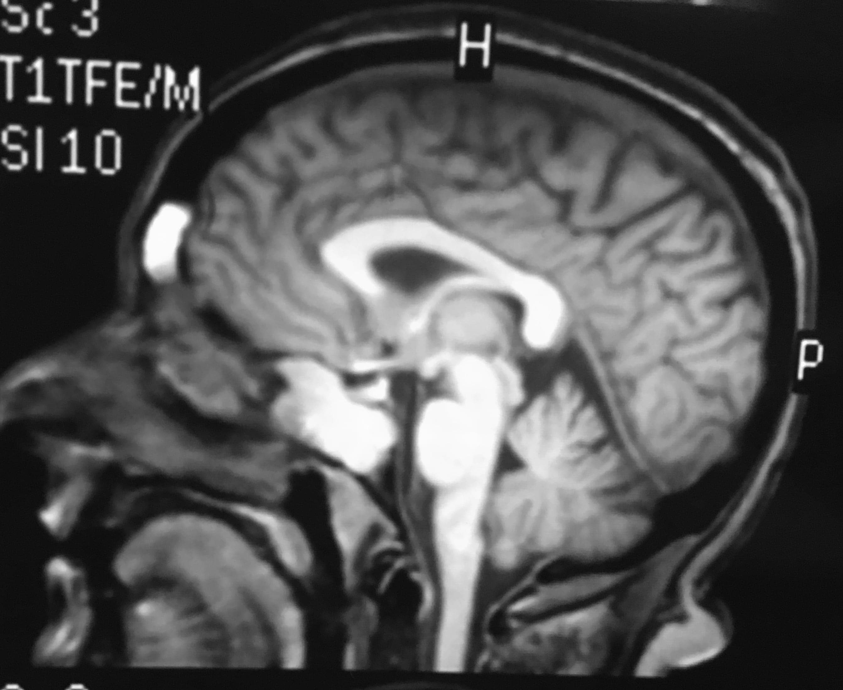
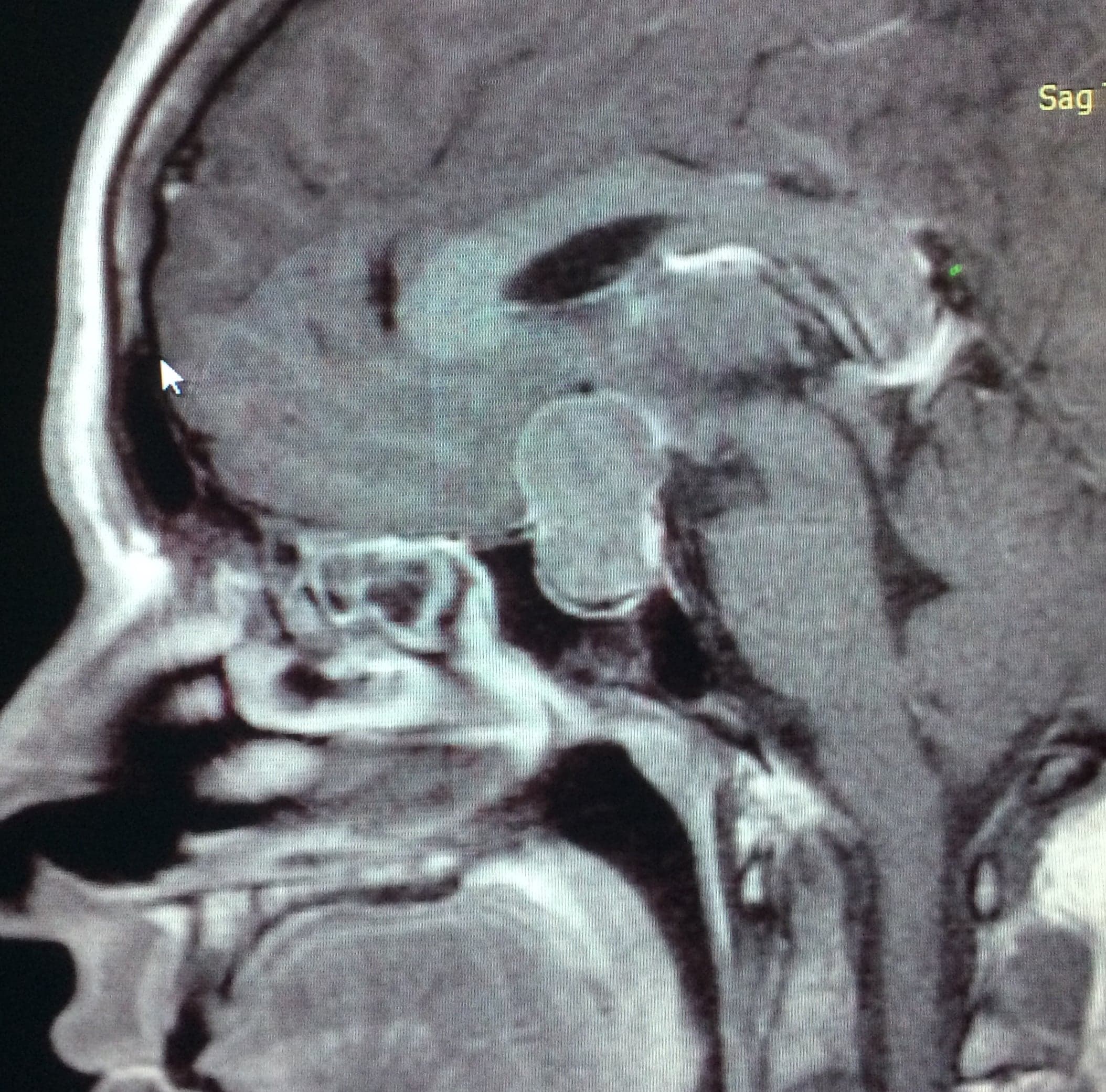
A 58-year-old patient consulted us for double vision and headaches. MRI showed a skull base lesion in the upper clivus that, in its intracranial growth, displaces the pituitary upward, erodes the bone, and lies anterior to the brainstem. Endoscopic endonasal surgery was performed with a good outcome and complete resection of the lesion. Pathology confirmed the diagnosis of Clival Chordoma.

As can be seen in the surgical video, endoscopic endonasal surgery allows resection of skull base tumors and provides high-definition images of normal brain structures essential for correct surgical resection.

Second Case

A 23-year-old patient was referred to our institution for a skull base tumor compromising the brainstem. MRI showed a tumor compatible with Clival Chordoma compressing and partially invading the brainstem. Clinically, the patient suffered from double vision and dysmetria in all four limbs, with gait alteration and impaired fine motor movements of the hands. Complete endoscopic resection was performed with a good outcome. The patient developed a CSF leak, which was resolved with a new endoscopic closure. Six months later, the patient fully recovered the neurological symptoms and can lead a completely normal life.

Third Case

Lastly, we want to illustrate our experience with a case of Giant Clival Chordoma. A 30-year-old patient came to consultation for chronic headache related to Valsalva maneuvers or exercise, hoarseness and swallowing disturbances. Imaging showed a giant skull base tumor with extensive intracranial extension and brainstem compression. Endoscopic endonasal surgery allows us to resect large skull base tumor volumes, since the tumor itself creates the space for resection, displacing noble structures such as cranial nerves and the brainstem. An extended endoscopic endonasal surgery was performed for the widest possible resection. Fortunately, we achieved a good resection without complications, as evidenced by the surgical video and the postoperative images. The patient had very good symptomatic recovery and continues with periodic imaging follow-up.

Today, skull base surgery is performed by teams of neurosurgeons trained in both conventional and endoscopic endonasal surgery, with the goal of making the best decisions and improving the quality of life of patients with these pathologies.

Related cases

Esthesioneuroblastoma or Olfactory Neuroblastoma

Esthesioneuroblastoma or Olfactory Neuroblastoma

Esthesioneuroblastoma is a rare malignant tumor of the anterior skull base originating from olfactory cells.

View clinical case
Pituitary Adenomas

Pituitary Adenomas

Benign, slow-growing tumors originating from cells of the adenohypophysis, classified as functioning or non-functioning.

View clinical case
Craniopharyngiomas

Craniopharyngiomas

Benign tumors of the sellar and suprasellar region derived from epithelial cells of Rathke's pouch.

View clinical case