Esthesioneuroblastoma or Olfactory Neuroblastoma
March 13, 2019Pituitary adenomas
March 16, 2019Craniopharyngiomas

Craniopharyngiomas
Craniopharyngiomas are benign tumors of the sellar and suprasellar region derived from the epithelial cells in Rathke´s pouch. Children and older adults have a higher incidence rate. The most common symptoms are: alteration of the field of vision, hormonal deficits and short stature in children. When these tumors generate hydrocephalus, they can cause headaches, vomiting and sensory alterations.
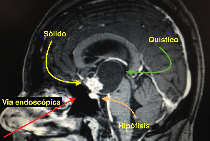
The presumptive diagnosis is made with an MRI and a Brain Tomography, where we can see the typical suprasellar, solid-cystic tumor with calcifications.
The best treatment for these tumors is total resection when possible. We have radiotherapy as an adjuvant for remnants of active tumors or for recurrent tumors.
Since the Craniopharyngiomas that originate in the hypothalamus pituitary axis are mainly midline and are in close relationship with the skull base, the endoscopic endonasal approach is the most frequently chosen path since it provides the most direct access to the region.
Our team prioritizes the endoscopic approach because it allows us to see the tumor more clearly and how it is positioned related to the hypothalamic pituitary axis in order to have the best chance of performing a total resection.
We present the case of a 20-year-old patient who experienced progressive loss of vision accompanied by drowsiness due to cortisol deficiency.
-

- We can observe a voluminous solid cystic tumor in the suprasellar cistern and in the third ventricle. Note that the endoscopic approach allows us to reach the tumor without having to mobilize the brain.
-

- In the coronal section we observed that the tumor was generating edema in the right hypothalamus. Compare this finding with intraoperative endoscopic vision imagine in which the bloody surface of the right hypothalamus is seen.
-

- After the dural opening we can clearly see the pituitary gland, the pituitary stem coming out of it and the tumor that is invading the upper part of the stem next to the floor of the third ventricle.
-

- Just after performing the total tumor resection we can see part of the normal brain, the roof of the third ventricle, which is tumor-free.
-

- Using angled optics we can see the interventricular foramina (or foramina of Monro) and the third tumor-free ventricle.
-

- If we rotate the view to the left side of the patient, we can see the left hypothalamus with no tumor and uninjured.
-

- Turning the view to the right, we see the right hypothalamus, which is bloody after the tumor resection since it is the most common site of adhesions. This image is consistent with the coronal section of the preoperative MRI.
-

- In this case, we were able to perform a total resection of the craniopharyngioma. The postoperative period was without complications. The correct total hormone replacement was performed and we did not need to use radiotherapy.
-

- Note the high definition of the intraoperative images provided by the endoscopic endonasal route. This view allows us to make the most appropriate decision during the surgery in order to obtain the best result possible.
In this case, we were able to perform a total resection of the craniopharyngioma. The postoperative period was without complications. The correct total hormone replacement was performed and we did not need to use radiotherapy.
Note the high definition of the intraoperative images provided by the endoscopic endonasal route. This view allows us to make the most appropriate decision during the surgery in order to obtain the best result possible.

